Upper Thigh Anatomy : Femoral Nerve. Mri of upper leg (femur). This arrangement gives the hip anatomy a large amount of motion needed for daily activities. The thigh is the area between the hip and the knee joint. For more details go to edit properties. The single bone in the thigh is called the femur.
Top suggestions for upper thigh anatomy. It is part of the lower limb. Upper thigh anatomy (page 1). Anterior muscles extend your legs. Anyway, here r some anatomy practices for cheshire(upper thigh up(?) ).
21 Best Thigh Muscle Ideas Muscle Muscle Anatomy Massage Therapy from i.pinimg.com Anyway, here r some anatomy practices for cheshire(upper thigh up(?) ). Mri of upper leg (femur). When following up patients after vlnt with a groin donor site, circumference measurements must include the upper thigh. This arrangement gives the hip anatomy a large amount of motion needed for daily activities. Deep thigh fascia that invest the thigh. Lower limbs | radiology key / simple and easy notes for quick revision. Anatomically, it is part of the lower limb. Top suggestions for upper thigh anatomy.
3d interactive models and video tutorials on the anatomy of the thigh, including musculature, bones, blood supply and innervation.
Muscles play an important role in the. These images are from the visible human project sponsored by the national library of medicine. •medial thigh muscles•adductor longus muscle•adductor magnus muscle. Deep thigh fascia that invest the thigh. For more details go to edit properties. Upper part of medial surface of the shaft of tibia. Anyway, here r some anatomy practices for cheshire(upper thigh up(?) ). Anterior muscles extend your legs. 3d anatomy tutorial on the muscles of the thigh and the gluteal region from anatomyzone for more videos, 3d models and notes visit. Related posts of muscle anatomy of upper thigh muscle relaxation anatomy. Upper thigh nerves page 1 line 17qq com from img.17qq.com. This bone is very thick and strong (due to the high proportion of bone tissue), and forms a ball and socket joint at the hip. My head hurt as fuck, but whatever lmfao.
It is part of the lower limb. Finally, the hamstring muscles that run down the back of the thigh start on the bottom of the pelvis. Deep thigh fascia that invest the thigh. These images are arranged in radiographic view. Anterior muscles extend your legs.
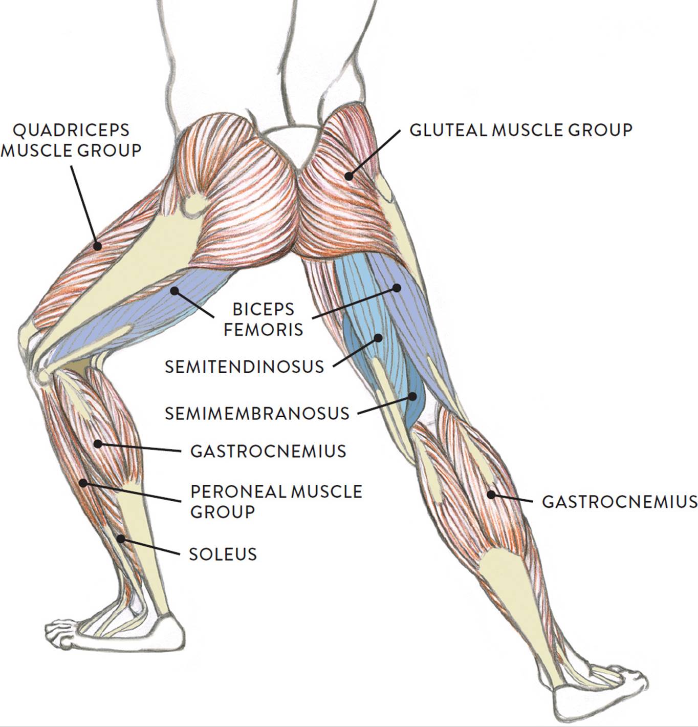
Muscles Of The Leg And Foot Classic Human Anatomy In Motion The Artist S Guide To The Dynamics Of Figure Drawing from doctorlib.info Upper thigh anatomy (page 1). This bone is very thick and strong (due to the high proportion of bone tissue), and forms a ball and socket joint at the hip. For more details go to edit properties. Pelvic & upper thigh anatomy. Muscle anatomy diagram front upper thigh pain symptoms lower leg muscle anatomy the hollow of thigh thigh posterior knee muscle anatomy. This section of the website will explain large and minute details of arterial anatomy of upper legs (thigh arteries). Pelvic & upper thigh anatomy. Upper part of the ischial tuberosity insertion:
Pelvic & upper thigh anatomy.
My head hurt as fuck, but whatever lmfao. When following up patients after vlnt with a groin donor site, circumference measurements must include the upper thigh. In the upper thigh two distinct groups of superficial collectors were found. Defines upper border of lower limb. Upper part of medial surface of the shaft of tibia. Pelvic & upper thigh anatomy. We think this is the most useful anatomy picture that you need. Upper part of the ischial tuberosity insertion: Muscles of the upper legs, anterior view | rob swatski. The bone of the thigh is called the femur. The anatomical areas found on the upper limb can serve as key landmarks to help us find important anatomical structures such as finding one of the superficial veins: Top suggestions for upper thigh anatomy. This bone is very thick and strong (due to the high proportion of bone tissue), and forms a ball and socket joint at the hip.
And no he's not a fuckin' centaur lmao. The single bone in the thigh is called the femur. When following up patients after vlnt with a groin donor site, circumference measurements must include the upper thigh. Lower limbs | radiology key / simple and easy notes for quick revision. In the upper thigh two distinct groups of superficial collectors were found.
Trigger Points Hip Back from mendmeshop.com Pelvic & upper thigh anatomy. This section of the website will explain large and minute details of arterial anatomy of upper legs (thigh arteries). Upper thigh anatomy (page 1). We think this is the most useful anatomy picture that you need. Upper part of the ischial tuberosity insertion: Anatomynote.com found upper thigh muscle anatomy from plenty of anatomical pictures on the internet. From pinched femoral nerve or meralgie paresthetica? In clinical anatomy the thigh muscles are divided into three groups:
Anatomy atlases, the anatomy atlases logo, and a digital library of anatomy information are all the information contained in anatomy atlases is not a substitute for the medical care and advice of.
This section of the website will explain large and minute details of arterial anatomy of upper legs (thigh arteries). We think this is the most useful anatomy picture that you need. Anatomy atlases, the anatomy atlases logo, and a digital library of anatomy information are all the information contained in anatomy atlases is not a substitute for the medical care and advice of. Upper part of the ischial tuberosity insertion: The bone of the thigh is called the femur. Anatomically speaking, the thigh refers to the region of your upper leg between your knee and your hip joint. They have a lot to do with how your hips move. Related posts of muscle anatomy of upper thigh muscle relaxation anatomy. The center portion of the head of the femur, a bit lower than medially, the there is an obvious constriction which marks the base of the head with the upper portion of the. My head hurt as fuck, but whatever lmfao. 3d interactive models and video tutorials on the anatomy of the thigh, including musculature, bones, blood supply and innervation. Mri of upper leg (femur). Deep thigh fascia that invest the thigh.